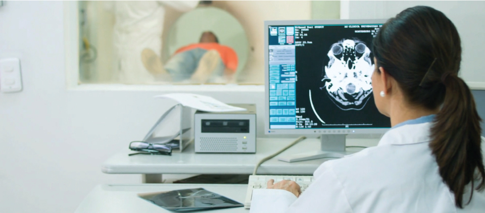
A computed tomography scan (better known as a “CT scan”) uses a narrow x-ray beam to take multiple measurements of the body. These images are then joined together by sophisticated computers, resulting in a cross-sectional image of the scanned object, which greatly helps with diagnoses and treatment.
Areas of Application
CT of the Head
CT brain, a CT scan of the head produces a cross-section of your brain and can provide detailed information on head injuries, stroke, brain tumors, and other brain diseases. This imaging technique details bone, soft tissues, and blood vessels.
Benefits
- Detection of bleeding, brain damage, and skull fractures in patients with head injuries.
- Detecting a blood clot or bleeding within the brain shortly after a patient exhibits symptoms of a stroke.
- Detection of stroke.
- Evaluation of the extent of bone and soft tissue damage in patients with facial trauma.
- Detection of bleeding in a patient with a sudden severe headache who may have a ruptured or a leaking aneurysm.
- Detection of most brain tumors.
- Diagnosing diseases of the temporal bones, which may be causing hearing/balance problems or tinnitus.
- Detection of enlarged brain cavities (ventricles) in patients with hydrocephalus.
- Determining whether infection/inflammation or other changes are present in the paranasal sinuses.
- Planning radiation therapy for cancer.
- Non-invasive assessment of aneurysms or arteriovenous malformations through a technique called CT Angiography.
- Detecting diseases or malformations of the skull.
- CT Angiography depicts blood vessels supplying the brain, revealing aneurysms and vessel narrowing/occlusion.
- Three-dimensional imaging of the skull and brain structures for surgical planning.
CT of the Chest
A CT scan of the chest is often used to further evaluate abnormalities found on routine chest x-rays. Because CT allows much greater clarity of internal organs, bone, soft tissue, and blood vessels than can be seen with routine x-rays, physicians are able to perform a more in-depth study.
Benefits
- Further evaluates abnormalities found on routine chest x-rays.
- Help diagnose the cause of symptoms such as a cough, shortness of breath, chest pain, or fever.
- Evaluate an injury to the chest.
- Detect infections such as pneumonia or tuberculosis.
- Detect lung cancer.
- Demonstrate chronic conditions such as emphysema, bronchiectasis or interstitial lung disease.
- Diagnose vascular abnormalities such as pulmonary emboli (blood clots in the lung vessels) or an aortic aneurysm (dilated aorta).
CT of the Body (Abdomen and Pelvis)
A CT scan of the body is typically performed on the abdominal and pelvic region. Because CT allows much greater clarity of internal organs, bone, soft tissue, and blood vessels, CT is very well suited for evaluation and diagnosis of numerous disorders that can occur in these regions.
- Diagnose infections such as diverticulitis, appendicitis, or pyelonephritis (kidney infection).
- Detect tumors in the liver, pancreas, or kidney.
- Find stones in the kidney or bladder.
- Evaluate enlarged lymph nodes in lymphoma or leukemia.
- Diagnose and follow an aortic aneurysm (enlarged aorta).
- Screen patients with abdominal trauma for organ injury.
- Rapid evaluation of patients with nonspecific abdominal pain.
Coronary CT Angiography
A Coronary CT Angiography (Coronary CTA) exam can help be done for several reasons, such as:
- To examine the thoracic aorta with coronary angiography
- For pulmonary vein mapping
- To show cardiac anatomic structure
- For valve replacement planning
We also offer Functional Flow Reserve analysis of coronary CTA.
HeartFlow® technology uses computed tomography (CT) scans to calculate how much blood is flowing through the coronary arteries. This test helps to identify whether patients have coronary artery disease before undergoing invasive and possibly unnecessary catheter-based testing.
FFR-CT helps guide treatment strategies and determines if blood flow is being restricted when flowing through an artery. FFR-CT is very low risk and provides accurate information about conditions of the arteries. It can determine whether a specific plaque in a coronary artery is restricting blood flow, thereby helping determine whether a patient would benefit from stents or bypass surgery.
FFR-CT can benefit patients who have experienced symptoms such as chest pain and shortness of breath, but have not been diagnosed with heart disease.
This analysis does not require a separate CT and can be ordered by a physician either before or after the exam has taken place.
CT Benefits vs Risks
Benefits
- CT scans are fast, non-invasive, and accurate.
- They are capable of imaging bone, soft tissue, and blood vessels all at the same time.
- A diagnosis determined by CT scanning may eliminate the need for exploratory surgery and biopsy.
- CT scans have been shown to be a cost-effective imaging tool for a wide range of clinical problems.
Risks
- Although the patient is exposed to clinically acceptable radiation, the benefit of an accurate diagnosis far outweighs the risk.
- Intravenous contrast material is often used when performing CT studies; the risk of serious reaction is very rare and our personnel is trained to screen all patients and treat any reaction.
- Women should always inform their doctor and CT technologist if there is any possibility that they are pregnant.
- Nursing mothers should wait 24 hours after contrast injection before resuming breastfeeding.
How Do CT Scans Work?
Unlike conventional x-rays, which produce one-dimensional images of the body, CT scanning uses x-rays in a much different way. During a CT scan, numerous x-ray beams are passed through the body at different angles, and special sensors measure the amount of radiation absorbed by different tissues.
As you lay still, the scanner revolves around you (although you cannot see this happen), emitting x-ray beams in a spiral path, while recording thousands of data points. This technique is called helical or spiral CT. A special computer program then analyzes the differences in x-ray absorption to form cross-sectional images, or “slices.”
CT imaging is sometimes compared to looking into a loaf of bread by cutting the loaf into thin slices or tomograms, hence the name “computed tomography.” The images can then be reconstructed in any two-dimensional plane or into three-dimensional images of the body’s interior.
How is a CT Scan Performed?
During a CT scan, a technologist will begin by positioning you on the CT examination table, usually lying flat on your back. The table will move slowly through the machine as the actual CT scanning is performed. You will be asked to hold your breath during the scanning. Any motion, whether breathing or body movements, can lead to artifacts on the images, so it is very important to follow instructions. Contrast material may be injected unless there is a history of allergy or other contraindications.
How to Prepare for Your Examination
Metal objects including jewelry, eyeglasses, dentures, dental implants or hairpins may adversely affect the CT images and should be left at home or removed prior to your exam.
You may be asked not to eat or drink anything for several hours beforehand, especially if a contrast material or “dye” will be used in your exam. You should inform your physician of any medications you are taking and if you have any allergies including allergy to contrast material, or “dye”. It is especially important to notify the staff if you have a history of heart disease, asthma, diabetes or kidney disease – particularly if you are taking Glucophage (Metformin) – as these conditions may affect your response to the contrast material.
Women should always inform the technologist and radiologist if there is any possibility that they are pregnant or nursing.
Who Will Interpret the CT Scan?
A radiologist with special expertise in the targeted area for the CT scan will analyze the images. Head CT scans will be interpreted by a neurologist. Chest CT examinations are interpreted by a radiologist experienced in chest CT examinations. Body CT scans are read by a radiologist with expertise in abdominal and pelvis CT examinations.
A preliminary interpretation may be available shortly after the exam. The completed report is sent to the referring physician within twenty-four hours. Your primary physician or the radiologist may discuss the findings of the CT examination with you. Mink Radiology also allows for distribution of diagnostic reports and images over the Internet from our facility to your doctor.
Next, learn about Ultrasounds.