

Mink Radiology
Has joined the RadNet/Cedars-Sinai family!

Patient Portal
ACCESS PORTALBILLING QUESTIONS
Studies Completed Before 9/1
Please call: 1-800-964-9651
Studies Completed After 9/1
Please call: 1-844-866-2718
IMAGING
SERVICES
OPEN MRI
Unlike a traditional MRI machine, an open MRI features open space on either side of the machine, which helps reduce and eliminate claustrophobia and/or anxiety. With an open MRI, patients can expect high-quality results in a more relaxing and comfortable environment.
MRI
An MRI allows us to produce extremely detailed pictures of body tissues and organs without the need for x-rays. During an MRI, electromagnetic energy is measured and analyzed to produce both two- and three-dimensional images that can be viewed on a computer monitor. This advanced imaging technology allows for the evaluation of some body structures that may not be as visible with other imaging methods.
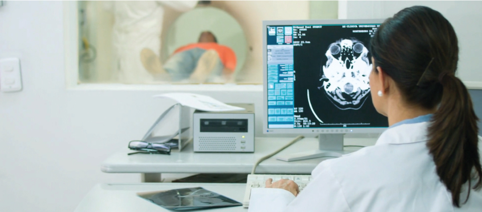
CT SCAN
A computed tomography scan (better known as a "CT scan") uses a narrow x-ray beam to take multiple measurements of the body. These images are then joined together by sophisticated computers, resulting in a cross-sectional image of the scanned object, which greatly helps with diagnoses and treatment.
ULTRASOUND
An ultrasound is a diagnostic imaging technique that uses sound waves to "see" inside the body. Unlike other imaging modalities (such as x-rays), ultrasounds capture live information, which means that our specialists can see the structure and the movement of the body's internal organs in real time.
X-RAY
X-rays are the oldest and most frequently used form of medical imaging. They are a form of electromagnetic radiation like light or radio waves. Imaging with x-rays involves exposing a part of the body to a small dose of ionizing radiation to produce pictures of the inside of the body.
BREAST IMAGING
At Mink Advanced Imaging, we specialize in a variety of imaging modalities. We offer breast imaging and mammography in comfortable, private, and discreet examination rooms. Other imaging modalities include radiography and x-rays, which are non-invasive and painless tests performed by our radiographic specialists.
OPEN MRI
Unlike a traditional MRI machine, an open MRI features open space on either side of the machine, which helps reduce and eliminate claustrophobia and/or anxiety. With an open MRI, patients can expect high-quality results in a more relaxing and comfortable environment.
MRI
An MRI allows us to produce extremely detailed pictures of body tissues and organs without the need for x-rays. During an MRI, electromagnetic energy is measured and analyzed to produce both two- and three-dimensional images that can be viewed on a computer monitor. This advanced imaging technology allows for the evaluation of some body structures that may not be as visible with other imaging methods.
CT SCAN
A computed tomography scan (better known as a "CT scan") uses a narrow x-ray beam to take multiple measurements of the body. These images are then joined together by sophisticated computers, resulting in a cross-sectional image of the scanned object, which greatly helps with diagnoses and treatment.
PAIN MANAGEMENT
Image guided procedures are minimally-invasive treatments that are commonly used for diagnostic and pain management purposes. The imaging experts at Mink Advanced Imaging perform joint injections (such as epidural and nerve blocks for the spine) and myelography, which provides a real-time look at the movement and structure of the spine.
DEXA BONE SCAN
A Bone Density Scan (DEXA) is a swift and non-invasive procedure that measures the density and mineral content in bone, primarily focusing on the hip or lower spine. This advanced imaging technique uses a low-dose x-ray to accurately assess bone health, aiding in the diagnosis and management of conditions such as osteoporosis.
CONTACT MINK ADVANCED IMAGING


